The post Nicole Kidman’s new role shines a light on genetics first appeared on Know Pathology Know Healthcare.
]]>Photograph 51 relates Franklin’s contribution to the discovery of the double helix structure of DNA in the 1950s. The play depicts the sometimes confrontational working relationship between the talented Franklin and her laboratory partner, Maurice Wilkins.
The play’s name comes from the X-ray image of DNA that Franklin created. It was this image that led scientists James Watson and Francis Crick to determine the chemical structure of DNA, ushering in the age of modern genetics.
In 1962, the Nobel Prize in Physiology or Medicine was awarded to Watson, Crick and Wilkins, with Franklin notably overlooked. In 1958, Franklin died of cancer, never having been recognised for her work. Photograph 51 attempts to bring Franklin’s role to light.
Dr Melody Caramins is a genetic pathologist working in Sydney. She says modern medicine would look very different without the discovery.
“Genetic testing is widely used, particularly for screening; for example prenatal testing for Down Syndrome and newborn bloodspot testing for life-threatening conditions like Cystic Fibrosis.
Genetic testing can also suggest if a particular cancer drug is likely to be effective for an individual patient. Testing can also indicate an elevated risk of developing a hereditary cancer.”
Dr Caramins says that genetics is an exciting and rapidly developing area to work in as there are so many questions to be answered.
“I encourage anyone willing to work hard to consider pathology and genetics in particular. There is great variety in the work on offer, including lab work and consulting directly with patients.”
This burgeoning profession owes much to genetic pioneers like Rosalind Franklin.
The post Nicole Kidman’s new role shines a light on genetics first appeared on Know Pathology Know Healthcare.
]]>The post “After nine years I still get a rush out of my work” – up close and personal with the people behind pathology first appeared on Know Pathology Know Healthcare.
]]>Medical Scientist – Alicia Thompson
Alicia had been interested in science since high school and after completing her studies went to work as a food scientist testing dairy products for safety. Alicia enjoyed the work but always knew she wanted to move into medical science;
“I liked the fact there was a human being at the end of a test”. Alicia said, “After nine years I still get a rush out of my work. It’s stressful when we’re really busy but I thrive off the adrenaline rush. The phone rings and it could be anything from a car accident with multiple traumas needing blood transfusions, or a critically ill baby needing a test for an infection around the brain. Every day you’re jumping into the unknown.”
When asked about memorable moments Alicia said:
“There are lots of small moments that have made me proud of my work over the years. Often if a patient is going into surgery I’ll try to prepare in advance for potential requests from the surgeons. A couple of times the patient was bleeding heavily and they needed more red blood cells. When I’ve been able to pre-empt the request from the surgeon and have everything ready to go immediately they’re very impressed. It’s not often we get a ‘thank you’ but on those occasions the anaesthetist has called me up directly to thank me for my role in helping to save a patient’s life. That feels pretty good!”
Pathologist – Dr David Clift
David says that during his medical training he was at first intending to become a surgeon.
“As an undergraduate student I didn’t understand the importance of pathology but as a resident in a hospital working for a cancer surgeon, and also working in a haematology department, I got to see how important and interesting pathology was.”
David says that his area of pathology is incredibly valuable and interesting. He enjoys the personal diagnostic challenges that are different every day but also knows the importance of being able to help another medical practitioner to provide a more personalised service. He says that pathologists also see rare conditions more often than many other practitioners, which helps them to provide better advice to surgeons.
One case of an uncommon disease that David diagnosed helped safeguard a group of children:
“One of the most dramatic cases where my work made a difference was when I made a diagnosis of a serious infectious disease at autopsy. It was meningococcal septicaemia in a young child; I made the diagnosis in the morning and public health measures were being taken by the afternoon for a whole grade one class. I have grandchildren at this age so it felt good to work together with microbiology and public health to help protect these children.”
Collection Services Manager – Peta Martin
Peta started as a lab technician in the Royal Australian Air Force and while working there trained as a medical scientist. Peta said that working as a medical scientist in the RAAF was different from her later roles in the civilian world;
“We were working with a closed population of people who were generally healthy so a lot of what we did was screening tests, but we also had hospitals and field medical units. We did everything from collection to ward rounds and working in the lab.”
The thing Peta likes most about her current role as a Collection Services Manager is the direct contact with patients. “We’re the face of pathology, the people the patient remembers,” she said.
A particular moment which is a good example of this personal touch happened during a time Peta worked in a country setting;
“We had a lady with a condition called cold agglutinins causing a haemolytic anaemia* – once the blood samples are collected, as soon as the blood starts to cool the red cells agglutinate (clump together) resulting in many tests being impossible to perform on those samples. Everything had to be warmed up for this patient; from the bleeding room to the tubes, the collection devices and needles. Watching my staff go out of their way to help this lady and knowing we were helping her in her crisis moment was pretty wonderful.”
*Hemolytic anemia is a condition in which red blood cells are destroyed and removed from the bloodstream before their normal lifespan is over
The post “After nine years I still get a rush out of my work” – up close and personal with the people behind pathology first appeared on Know Pathology Know Healthcare.
]]>The post Analyse this: so what exactly goes into diagnosing coeliac disease? first appeared on Know Pathology Know Healthcare.
]]>Many people with coeliac disease know that getting a confirmed diagnosis can be a long process, so how exactly is this done, and who is responsible for getting the test results?
18th November 2015 is International Pathology Day so why not sign up and tell us your coeliac disease diagnosis story via the comment box on our homepage
When a doctor suspects coeliac disease, the first pathology test that is usually ordered is coeliac serology. Serology testing looks for the presence of specific antibodies in a person’s blood. Antibodies are normally the ‘good guys’ – they attack infections. In autoimmune conditions like coeliac disease, autoantibodies form, which mistakenly attack the body’s own organs. The coeliac serology tests look for these autoantibodies which attack the small bowel (small intestine) as part of an abnormal reaction to gluten.
The autoantibodies are found in serum – the watery component of blood in which red and white blood cells float as they pass through the body. To obtain serum for testing, the blood tube is sent to the pathology laboratory where it is spun in a centrifuge at 3000 revolutions per minutes. That’s two to three times faster than your washing machine on fastest spin. Doing this separates the yellow serum from the heavier red blood cells, which sink to the bottom of the tube.
Once the tube of blood has been spun it is loaded onto a machine where a pipette takes a few drops of the serum and adds them to reaction chambers (small plastic or glass disposable vials) containing various chemicals. Each reaction chamber will produce a specific reaction if the blood contains the autoantibodies that the test is looking for.
This process is overseen by a highly trained medical scientist who will interpret the results from the machine before they are delivered to the patient’s doctor.

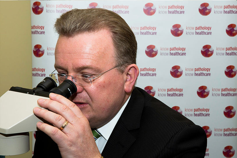
MP Bruce Billson, who has been diagnosed with coeliac disease, visited a pathology laboratory earlier this year and showed his support for the Know Pathology Know Healthcare campaign
Positive serology results show the person’s body is producing an inappropriate immune response, but for a confirmed diagnosis of coeliac disease, a biopsy is needed.
The patient is given a light anaesthetic and a small sample of tissue is collected from the lining of the small bowel. The tissue sample is typically only around 3mm in diameter.
The specimen is put into tissue fixative substance, usually formalin and sent to the pathology laboratory.
A pathologist or medical scientist then performs what’s known as a gross examination – this means looking at the sample without a microscope. This is done to record what can be seen by simply looking at, measuring, or feeling the tissue, for example its size, colour and consistency.
The sample is then processed and placed in hot paraffin wax, which sets as a solid block with the tissue within it.
Next, a microtome instrument is used to cut the block into very thin slices. The slices need to be thin enough to let light through in order for cells to be seen under a microscope. Slices are placed on glass slides, which are dipped into different stains or dyes that change the colour of the tissue to make cells more visible under a microscope.
The pathologist then examines the slides, looking for changes consistent with coeliac disease. When viewed under a microscope, healthy bowel tissue features small ‘finger-like’ projections called villi. These are typically shortened in coeliac patients. The pathologist looks for the extent of any shortening and for the depths of the ‘crypts’ (the indents between villi) to see if these have changed.
The pathologist will also look for the presence of lymphocytes, a type of white blood cell, in the upper layer of the tissue and for increased inflammatory cells.
Once the review is complete, the pathologist prepares a report containing their findings for the patient’s doctor. It is important that the doctor has the information from both the blood test and the biopsy combined with the patient’s history in order to make a confident diagnosis of coeliac disease.
Pathology is clearly vital for people with coeliac disease. Tell us your coeliac disease story and why you value pathology, by signing up here www.knowpathology.com.au
The post Analyse this: so what exactly goes into diagnosing coeliac disease? first appeared on Know Pathology Know Healthcare.
]]>